
Critical medical emergencies for nurses. Learn key interventions for hypertensive encephalopathy, septic shock, MI, Cauda equina syndrome, herniated disc, atrial fibrillation, cardiac tamponade, testicular torsion. Master the skills needed to ace the exam and confidently respond to urgent clinical situations. Elevate your nursing career with this concise guide to passing the NCLEX-RN.”
- HYPERTENSIVE ENCEPHALOPATHY
- SEPTIC SHOCK
- MYOCARDIAL INFARCTION
- CAUDA EQUINA SYNDROME
- HERNIATED DISC
- ATRIAL FIBRILLATION
- CARDIAC TAMPONADE
- TESTICULAR TORSION
HYPERTENSIVE ENCEPHALOPATHY:
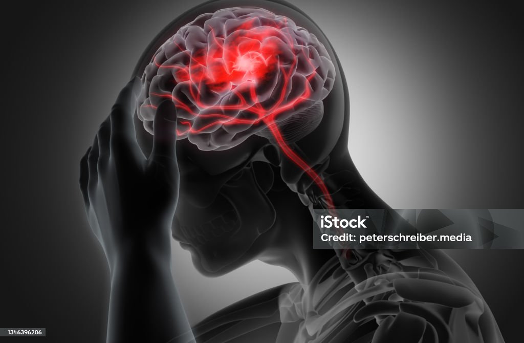
Hypertensive encephalopathy is a medical emergency characterized by rapidly increasing high blood pressure (hypertension) that leads to swelling and dysfunction in the brain.
This condition typically manifests as a spectrum of neurological symptoms, including headache, confusion, seizures, visual disturbances, and altered mental status.
If left untreated, hypertensive encephalopathy can progress to more severe complications, such as cerebral edema, intracranial hemorrhage, and brain damage.
Prompt recognition and management are essential to prevent further neurological deterioration and potential long-term consequences.
SEPTIC SHOCK
- Massive infection leads to sepsis as a result of the release of endotoxins from bacteria which further result in vasodilation and pooling the blood.
- Sign and symptoms: Persistent hypotension despite adequate fluid resuscitation requiring vasopressors, along with decreased tissue perfusion that progresses to tissue hypoxia.
- Three major effects of septic shock include vasodilation, maldistribution of blood flow, and myocardial depression.
- Clients who develop septic shock are at risk for the development of respiratory failure and acute respiratory distress syndrome.
- ROLE OF NURSE:
- Fluid replacement: 30-50 mL/kg fluid resuscitation with isotonic crystalloids, albumin may also be administered.
- Vasopressor therapy to increase cardiac output
- IV corticosteroids may be administered in clients
- IV antibiotics should be started within the first hours of sepsis or septic shock; cultures should be obtained before initiation of antibiotics therapy.
MYOCARDIAL INFARCTION
It occurs when myocardial tissue is abruptly and severe deprived of oxygen. Ischemia can lead to necrosis of myocardial tissue if blood flow is not restored. It does not occur instantly but evolves over several hours.
- Obvious physical changes do not occur in the heart until 6 hours after the infarction, when the infarcted area appears blue and swollen.
- After 48 hours, the infarct turns gray, with yellow streaks developing as neutrophils invade the tissue.
- By 8 to 10 days after infarction, granulation tissue forms.
- Over 2 to 3 months, the necrotic area develops onto a scar, scar tissue permanently changes the size and shape of the ventricle.
- ASSESSMENT:
- Pain:
- crushing substernal pain
- Radiate to the jaw, back, and left arm
- Pain may occur, primarily early in the morning
- Pain is unrelieved by rest or nitroglycerin and is relieved by only opioids.
- Pain lasts 30 minutes or longer
- Nausea and vomiting
- Diaphoresis
- Dyspnea
- Dysrhythmias
- Feeling of fear and anxiety
- Impending doom
- Pallor
- Cyanosis
- Coolness of extremities
- Pain:
- ROLE OF NURSE:
- Pain relief increases oxygen supply to the myocardium; administer MORPHINE as a priority in managing pain in the client with MI
- Administer OXYGEN and MORPHINE/ NITROGLYCERIN as prescribed
- Assess vital signs and Cardiovascular status.
- Maintain Cardiac monitoring.
- Assess respiratory rate and breath sounds for signs of heart failure (crackles, wheezes)
- Bed rest, Semi fowler position
- Stay with the client.
- Obtain a 12 lead ECG.
- Monitor laboratory values.
- Monitor for cardiac dysrhythmias because tachycardia and PVCs frequently occur in the first few hours after MI; administer antidysrhythmics as prescribed.
- Administer thrombolytic therapy (may be prescribed within the first hours of the coronary event).
- Monitor for signs of bleeding if the client is receiving thrombolytic therapy
- Assess distal peripheral pulses and skin temperature
- Monitor BP closely
- Provide reassurance to client and family.
Here is a mnemonic to remember the description “ONAM for Myocardial Infarction Treatment”:
O – Oxygen: Administering oxygen to the patient is crucial to increase the oxygen supply to the heart muscle and reduce the workload of the heart.
N – Nitroglycerin: Nitroglycerin helps dilate the blood vessels and improve blood flow to the heart. It is commonly used in the treatment of myocardial infarction.
A – Aspirin: Aspirin is given to patients to prevent blood clot formation by inhibiting platelet aggregation, thereby decreasing the risk of further damage to the heart.
M – Morphine: Morphine is used to relieve severe chest pain (angina) and reduce anxiety in patients with myocardial infarction.
CAUDA EQUINA SYNDROME
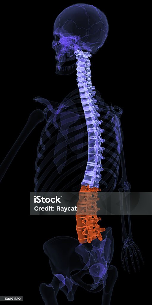
Occurs from injury to the lumbosacral nerve roots below the conus medullaris. The client experiences areflexia of the bowel, bladder, and lower reflexes.
Cauda Equina Syndrome (CES) is a medical emergency that occurs when the bundle of nerves at the base of the spinal cord, known as the cauda equina, is compressed. The cauda equina is responsible for transmitting signals to and from the lower limbs and pelvic organs. When compression occurs, it can lead to a range of symptoms that require immediate medical attention.
Common causes of Cauda Equina Syndrome include:
- Herniated Disc: When the disc material between vertebrae ruptures and presses on the nerves.
- Spinal Tumors: Abnormal growths in or around the spinal cord can cause compression.
- Spinal Infections: Infections affecting the spinal canal can lead to inflammation and compression of the nerves.
- Trauma: Severe injuries to the lower spine can result in CES.
Symptoms of Cauda Equina Syndrome may include:
- Severe Lower Back Pain: Often localized to the lumbar or sacral regions.
- Leg Weakness or Numbness: Difficulty moving or feeling the legs.
- Loss of Bladder or Bowel Control: Incontinence or difficulty urinating.
- Saddle Anesthesia: Numbness in the area that would come into contact with a saddle, affecting the inner thighs, buttocks, and genital area.
- Sexual Dysfunction: Loss of sensation or function in the genital area.
If you suspect Cauda Equina Syndrome, it is crucial to seek immediate medical attention. Early diagnosis and treatment are essential to prevent permanent neurological damage. Treatment may involve surgical intervention to relieve the pressure on the nerves.
Remember that the information provided here is for general knowledge, and it’s important to consult with healthcare professionals for accurate diagnosis and appropriate treatment options tailored to individual cases.
HERNIATED DISC:
A herniated disc, also known as a slipped disc or disc prolapse, occurs when the soft inner material of a spinal disc protrudes through the tough outer layer. Spinal discs are rubbery cushions between the vertebrae that act as shock absorbers for the spine. When a disc herniates, it can irritate nearby nerves, leading to pain, numbness, or weakness in the affected area.
Causes:
- Age: Discs naturally degenerate over time, becoming less flexible and more prone to herniation.
- Trauma: Sudden force or injury to the spine can cause a disc to herniate.
- Repetitive strain: Activities or occupations that involve repetitive bending, lifting, or twisting can increase the risk.
- Genetics: Some people may inherit a predisposition to disc problems.
Symptoms:
- Back or neck pain: The location and severity of pain can vary depending on the affected disc and the extent of nerve irritation.
- Radiating pain: Pain may radiate into the arms or legs if the herniated disc compresses spinal nerves.
- Numbness or tingling: Sensations of numbness, tingling, or pins-and-needles may occur in the arms or legs.
- Muscle weakness: Weakness in certain muscles, particularly those controlled by the affected nerves, may develop.
Diagnosis:
- Medical history: The doctor will inquire about symptoms, previous injuries, and medical history.
- Physical examination: This may involve testing reflexes, muscle strength, and sensation in different areas of the body.
- Imaging tests: X-rays, MRI (Magnetic Resonance Imaging), or CT (Computed Tomography) scans can provide detailed images of the spine to confirm the diagnosis and assess the extent of disc herniation.
Treatment:
- Conservative treatment: Initially, nonsurgical options such as rest, pain medications, anti-inflammatory drugs, physical therapy, and hot/cold therapy may be recommended.
- Epidural steroid injections: Injections of corticosteroids into the affected area can reduce inflammation and alleviate symptoms.
- Surgery: If conservative treatments fail to provide relief or if there are signs of severe nerve compression or neurological deficits, surgery such as discectomy or microdiscectomy may be considered to remove the herniated portion of the disc and relieve pressure on the nerves.
It’s essential to consult with a healthcare professional for proper diagnosis and treatment recommendations tailored to individual needs. Prompt medical attention can help manage symptoms effectively and prevent complications.
ATRIAL FIBRILLATION

Atrial fibrillation (AFib) is a common and potentially serious heart rhythm disorder characterized by irregular and often rapid heartbeats. In atrial fibrillation, the heart’s upper chambers (atria) beat chaotically and don’t effectively pump blood into the lower chambers (ventricles). This irregular pumping can lead to blood pooling in the atria, forming clots that may travel to the brain and cause a stroke.
Key points about atrial fibrillation:
Causes:
- Age: AFib is more common in older adults.
- Heart conditions: Conditions such as high blood pressure, coronary artery disease, and heart valve disorders can increase the risk.
- Other chronic conditions: Diabetes, obesity, and chronic kidney disease can contribute.
- Alcohol consumption: Excessive alcohol intake may increase the risk.
- Family history: A family history of atrial fibrillation can be a risk factor.
Symptoms:
- Irregular heartbeat: The most common symptom is a rapid, irregular heartbeat.
- Heart palpitations: Feeling of fluttering or thumping in the chest.
- Fatigue: Tiredness or weakness may be experienced.
- Shortness of breath: Difficulty breathing or a feeling of breathlessness.
- Dizziness or lightheadedness: Some individuals may feel dizzy or faint.
Complications:
- Stroke: AFib increases the risk of blood clots, which can lead to strokes.
- Heart failure: The irregular heart rhythm can lead to a weakened heart over time.
- Chronic fatigue: Persistent tiredness and reduced ability to exercise.
- Cognitive impairment: In severe cases, reduced blood flow to the brain may cause cognitive issues.
Diagnosis:
- Electrocardiogram (ECG or EKG): This test records the heart’s electrical activity and can detect irregularities.
- Holter monitor: A portable ECG device worn for a longer period to monitor heart activity.
- Blood tests: Checking for thyroid function and other factors.
- Echocardiogram: Using sound waves to create a detailed image of the heart’s structure and function.
Treatment:
- Medications: Anti-arrhythmic drugs, anticoagulants (blood thinners), and rate-control medications may be prescribed.
- Cardioversion: Electrical or chemical cardioversion may be used to restore normal heart rhythm.
- Ablation: Catheter ablation procedures can be performed to correct abnormal electrical pathways in the heart.
- Implantable devices: Pacemakers or implantable cardioverter-defibrillators (ICDs) may be recommended in some cases.
Management and treatment depend on the severity of symptoms, the underlying cause, and individual health considerations. It’s important for individuals with atrial fibrillation to work closely with their healthcare team to develop an appropriate treatment plan and reduce the risk of complications.
CARDIAC TAMPONADE
Cardiac tamponade is a medical emergency that occurs when fluid accumulates in the pericardial sac, the membrane surrounding the heart, and compresses the heart. This compression impairs the heart’s ability to pump blood effectively, leading to decreased cardiac output and potentially life-threatening complications.
Key points about cardiac tamponade:
Causes:
- Trauma: Penetrating or blunt chest trauma can lead to the accumulation of blood in the pericardial sac.
- Medical procedures: Complications from cardiac surgery, catheter placement, or percutaneous interventions can cause bleeding into the pericardial sac.
- Pericardial effusion: A buildup of fluid due to inflammation, infection, cancer, or other medical conditions can lead to tamponade if the fluid accumulates rapidly.
- Aortic dissection: A tear in the aorta can cause blood to leak into the pericardial sac and compress the heart.
Symptoms:
- Beck’s triad: Classic signs include hypotension (low blood pressure), muffled heart sounds, and jugular venous distention (enlarged neck veins).
- Dyspnea: Difficulty breathing due to decreased cardiac output.
- Tachycardia: Rapid heart rate as the heart attempts to compensate for decreased cardiac output.
- Pulsus paradoxus: An abnormally large drop in blood pressure during inspiration.
- Chest pain: Dull, aching chest discomfort may occur due to pressure on the heart.
Diagnosis:
- Physical examination: Clinical signs such as hypotension, jugular venous distention, and muffled heart sounds may suggest tamponade.
- Imaging tests: Echocardiography (ultrasound of the heart) is the primary diagnostic tool to visualize pericardial effusion and assess cardiac function.
- Electrocardiogram (ECG): ECG may show electrical alternans, a beat-to-beat variation in the QRS complex amplitude caused by swinging of the heart within the fluid-filled pericardial sac.
Treatment:
- Pericardiocentesis: Emergency drainage of fluid from the pericardial sac using a needle or catheter to relieve pressure on the heart.
- Fluid resuscitation: Intravenous fluids may be administered to support blood pressure and cardiac output.
- Inotropic medications: Drugs that increase cardiac contractility (e.g., dobutamine) may be used to improve heart function.
- Surgical intervention: In cases of recurrent tamponade or when pericardiocentesis is ineffective, surgical procedures such as pericardial window or pericardiectomy may be necessary to drain fluid and prevent recurrence.
Prompt recognition and treatment of cardiac tamponade are essential to prevent hemodynamic collapse and improve outcomes. It requires immediate medical attention and intervention to relieve the pressure on the heart and restore adequate cardiac function.
TESTICULAR TORSION
Testicular torsion is a medical emergency that occurs when the spermatic cord, which provides blood flow to the testicle, becomes twisted. This twisting can lead to a reduced or cut off blood supply to the testicle, causing severe pain and potential damage to the testicular tissue. Testicular torsion is most common in young males, often between puberty and the age of 25, but it can occur at any age.
Key points about testicular torsion:
Causes:
- Spontaneous torsion: The exact cause is often unknown, but it may be related to the mobility of the testicle within the scrotum.
- Physical activity or trauma: Strenuous physical activity, trauma to the groin, or sports injuries can contribute.
- Anatomical factors: Some individuals may have a predisposition to testicular torsion due to certain anatomical features.
Symptoms:
- Sudden, severe pain: Testicular torsion typically presents with rapid and intense pain in one testicle.
- Swelling: The affected testicle may become swollen and tender.
- Change in position: The testicle may appear higher than usual in the scrotum.
- Nausea and vomiting: These symptoms may occur due to the severe pain.
Diagnosis:
- Clinical examination: A healthcare professional will conduct a physical examination to assess symptoms and testicular position.
- Ultrasound: Imaging may be used to confirm the diagnosis and assess blood flow to the testicle.
Treatment:
- Manual detorsion: In some cases, a healthcare professional may attempt to manually untwist the spermatic cord to restore blood flow.
- Surgery (orchiopexy): Surgical intervention is usually required to permanently fixate the testicle and prevent future episodes of torsion. This procedure is called orchiopexy.
- Pain management: Pain relief medications and anti-inflammatory drugs may be prescribed.
Testicular torsion is a surgical emergency, and immediate medical attention is crucial to prevent irreversible damage to the testicle. If someone experiences sudden and severe testicular pain, they should seek medical help promptly. Delays in diagnosis and treatment can result in loss of the affected testicle and potential complications.
It’s important to note that not all cases of scrotal pain are due to testicular torsion, and other conditions such as epididymitis or trauma can cause similar symptoms. A healthcare professional will evaluate the individual’s symptoms and conduct appropriate tests for an accurate diagnosis.
Professional Nurse Practitioners: Top Nursing Programs for NP 2024 – brandednurses
1,000+ RN Jobs, Employment in Ontario 24 July 2024| Indeed.com